UOJM Volume 16, Issue 1
CASE REPORT
Misdirected eyelashes in a four-year-old girl with photophobia: Double the Trouble
2 Department of Ophthalmology, Faculty of Medicine, University of Ottawa, Ottawa, ON, Canada
3 University of Ottawa Eye Institute, The Ottawa Hospital, Ottawa, ON, Canada
4 Children’s Hospital of Eastern Ontario, Ottawa, ON, Canada
University of Ottawa Journal of Medicine, Volume 16, Issue 1, July 2026, pg. 32-35,
https://doi.org/10.18192/UOJM.V16i1.7515
Keywords: distichiasis; congenital eyelash disorder; photophobia; delayed diagnosis.
Abstract
One in ten thousand people are affected by distichiasis, a rare ocular abnormality whereby accessory eyelashes arise from or slightly posterior to the meibomian gland orifices. Given the non-specific symptoms, diagnosis is often missed or delayed, which can lead to corneal damage and compromise visual development in children. A 4-year-old girl was referred for longstanding, severe photophobia. Visual acuity was reduced (Snellen acuity 6/18 each eye). Gross inspection of the eyelid margin revealed an extra row of eyelashes emanating from the meibomian gland orifices in both the upper and lower eyelids bilaterally. Severe photophobia and diffuse bilateral punctate corneal epithelial erosions were also noted. The patient was diagnosed with congenital distichiasis which was repaired surgically using an eyelid-splitting technique with a combination of cryotherapy and radiofrequency ablation of the accessory eyelashes. This case emphasizes the importance of gross inspection of the external ocular structures in children presenting with persistent ocular symptoms.
Résumé
Une personne parmi dix mille est atteinte de distichiasis, une anomalie oculaire rare caractérisée par la présence de cils accessoires poussant au niveau ou légèrement postérieur des orifices des glandes de Meibomius. Compte tenu des symptômes non spécifiques, le diagnostic est souvent manqué ou retardé, ce qui peut entraîner des lésions cornéennes et compromettre le développement visuel chez les enfants. Une fillette de 4 ans a été orientée en raison d’une photophobie sévère et de longue date. L’acuité visuelle était réduite (selon l’échelle de Snellen de 6/18 pour chaque œil). L’examen macroscopique du bord palpébral a révélé une rangée supplémentaire de cils émanant des orifices des glandes de Meibomius, au niveau des paupières supérieures et des paupières inférieures, de manière bilatérale. Une photophobie sévère et des érosions épithéliales cornéennes ponctuées diffuses bilatérales ont également été observées. La patiente a reçu un diagnostic de distichiasis congénital, qui a été corrigé chirurgicalement à l’aide d’une technique de fendage palpébral en combinaison avec la cryothérapie et ablation par radiofréquence des cils accessoires. Ce cas souligne l’importance de l’examen macroscopique des structures oculaires externes chez les enfants présentant des symptômes oculaires persistants.
Case Presentation
A 4-year-old girl was referred to Paediatric Ophthalmology for severe photophobia from infancy, with a concern for possible albinism. The referring optometrist noted that the physical examination was limited, with the patient adopting a chin-down head position and maintaining closed eyelids during the exam; but refractive error, ptosis, and strabismus were all listed as findings in the referral. The family reported a ‘droopy left eyelid’ at birth; however, initial assessment by a paediatrician revealed no ocular abnormalities. A subsequent finding of reduced visual acuity in the right eye was treated with spectacle correction, but these were discontinued soon after, as the patient refused them. She complained of ocular pain and the family described mild yellow ocular discharge in the mornings. Symptoms improved with cold compresses. The family denied any history of pruritis, diplopia, flashes, floaters, or systemic symptoms including rashes or recent upper respiratory tract infections. Family history was non-contributory other than for a paternal grandfather with a ‘lazy eye’. At presentation, the patient’s visual acuity was below normal for age at 6/18 (Snellen) in each eye. Gross inspection of the eyelid margin revealed an extra row of eyelashes emanating from the meibomian gland orifices in both the upper and lower eyelids bilaterally (Figure 1A and 1B), with ocular surface irritation manifesting as diffuse bilateral punctate corneal epithelial erosions and reduced eyelid opening (appearing as a left upper eyelid ptosis). The patient was diagnosed with congenital distichiasis which was surgically repaired via eye-splitting with combined cryotherapy and radiofrequency eyelash ablation, with symptom resolution following surgery.
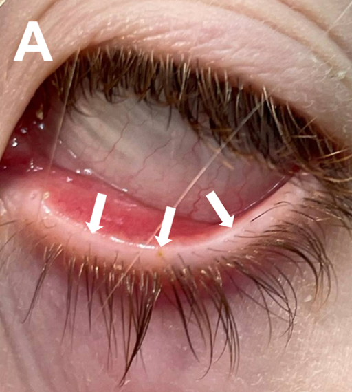
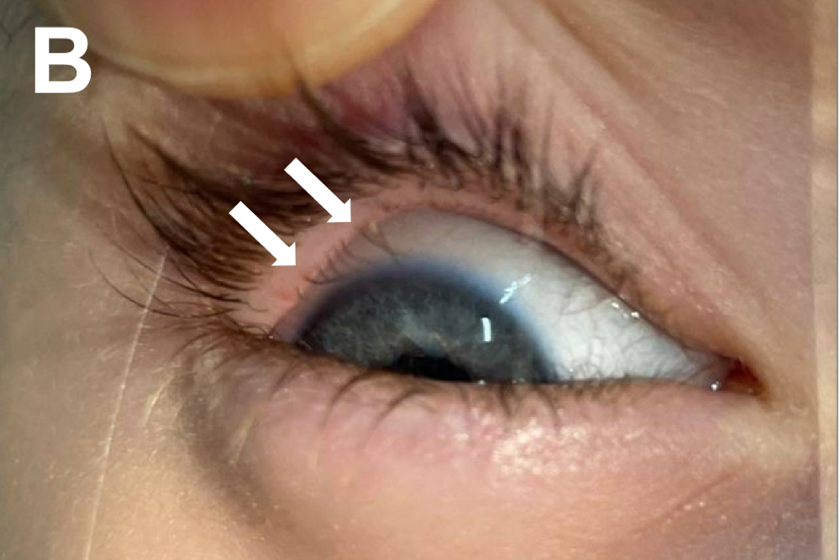
Figure 1. Patient’s (A) left and (B) right eye demonstrating accessory lashes arising from and slightly posterior to the meibomian gland orifices on the (A) lower and (B) upper eyelids (arrows).
Discussion
In this case, careful yet simple inspection of the eyelid margins revealed subtle aberrant lashes, leading to a rare diagnosis. Distichiasis is a rare ocular abnormality that affects 1 in 10,000 people whereby accessory eyelashes arise from or near the meibomian gland orifices.1,2 These glands are modified sebaceous glands located in the tarsal plate of the eyelids, which secrete meibum, an oil-rich substance, onto the ocular surface.3 Congenital distichiasis occurs when pilosebaceous units differentiate into eyelashes instead of meibomian glands, and may be observed in genetic syndromes including lymphedema-distichiasis syndrome and focal facial dermal dysplasia.2,4
Aberrant eyelashes can form a partial or complete secondary row on the upper and/or lower eyelids.4 During infancy, accessory eyelashes are typically soft and well-tolerated.5 However, they become problematic as they mature and irritate the cornea, causing pain and photophobia.2,4 If left untreated, this can lead to corneal epithelial defects, scarring, and astigmatism.6 Treatment is variable and often tailored to individual cases, largely dependent on location and extent of involvement.2 Multiple modalities such as electrolysis, cryotherapy, laser ablation, and surgical excision have been described in the literature with an overall success rate of 69-88%.2
While tempting for the provider and patient, eyelash epilation is counterproductive, as short, stubby eyelashes quickly regrow and inevitably cause worse corneal irritation than their predecessors.2,7 A series of 24 patients with distichiasis evaluated the treatment modalities of epilation, lid margin cryotherapy, and eyelid splitting cryotherapy to the posterior lamella and found that the latter effectively relieved symptoms without re-treatment in 87% patients.1 In severe bilateral cases, such as our patient, surgical eyelid-splitting with cryotherapy in a staged approach, supplemented by targeted radiofrequency ablation of eyelash follicles, under general anaesthesia is favoured.2
Distichiasis should be differentiated from other eyelash disorders which can cause similar symptomatology (Table 1). Trichiasis refers to normally-placed lashes which grow posteriorly towards the cornea, often secondary to inflammation and scarring of the eyelash follicles.8 Causes of trichiasis include chronic inflammatory disorders of the eyelid margin (blepharitis and meibomitis), dermatological conditions (atopic dermatitis, actinic elastosis and herpes zoster), conjunctival diseases (cicatricial trachoma and ocular pemphigoid), or eyelid margin scarring due to chemical, physical, or surgical trauma.8 Entropion involves inward rotation of the eyelid margin causing normal eyelashes to be misdirected posteriorly towards the corneal surface, resulting from involutional, mechanical (mass lesion), spastic (orbicularis muscle spasm), or cicatricial (conjunctival inflammation and scarring) changes.5,9 In the paediatric population, congenital defects and facial nerve palsy can also result in entropion.10 Epiblepharon, a congenital ocular condition common in people of Asian descent, involves the presence of a horizontal skin fold across the margin of the eyelid leading to vertical/inward positioning of the cilia, with potential for irritation and tearing.11
Table 1. Summary of differential diagnoses for distichiasis
| Diagnosis | Definition |
|---|---|
| Distichiasis | Accessory eyelashes arise from or near the meibomian gland orifices |
| Trichiasis | Normally-placed lashes which grow posteriorly towards the cornea |
| Entropion | Inward rotation of the eyelid margin causing normal eyelashes to be misdirected posteriorly towards the corneal surface |
| Epiblepharon | Presence of a horizontal skin fold across the margin of the eyelid leading to vertical/inward positioning of the cilia |
Slit lamp examination can provide a detailed assessment of the eyelid margin, skin, conjunctiva, cornea and direction of eyelash growth in cooperative patients.4 However, as seen in the case of our patient, gross inspection of the eyelids can identify misdirected or accessory eyelashes and also reveal scarring of the eyelid secondary to injury or chronic inflammation. Prompt identification can help to prevent delayed diagnosis and facilitate timely appropriate referral for adequate management.
Overall, this case emphasizes the importance of maximizing the use of gross inspection of external ocular structures in children. A high index of suspicion for rare external ocular abnormalities should be maintained for children presenting with persistent ocular symptoms. Diligent inspection of the eyelid margins in children with unexplained photophobia can prevent diagnostic delays and facilitate prompt referral for definitive management.
Conflicts of Interest Disclosure
There are no conflicts of interest to declare.
Informed Consent
Written informed consent was obtained from the patient’s family for
publication of this case.
References
1. O’Donnell BA, Collin JR. Distichiasis: management with cryotherapy to the posterior lamella. Br J Ophthalmol. 1993 May 1;77(5):289–92. doi:10.1136/bjo.77.5.289
2. Singh S. Distichiasis: An update on etiology, treatment and outcomes. Indian J Ophthalmol. 2022 Apr;70(4):1100–6. doi:10.4103/ijo.IJO_1141_21 PubMed PMID: 35325995; PubMed Central PMCID: PMC9240497.
3. Wu CY, Wu M, Huang X, Gu BJ, Maldonado-Codina C, Morgan PB, et al. Topographical Distribution and Phenotype of Resident Meibomian Gland Orifice Immune Cells (MOICs) in Mice and the Effects of Topical Benzalkonium Chloride (BAK). Int J Mol Sci. 2022 Aug 24;23(17):9589. doi:10.3390/ijms23179589
4. Patel BC, Joos ZP. Diseases of the Eyelashes. In: StatPearls [Internet]. Treasure Island (FL): StatPearls Publishing; 2025 [cited 2025 Mar 11]. Available from: http://www.ncbi.nlm.nih.gov/books/NBK537100/ PubMed PMID: 30725785.
5. Bowling B. Kanski’s clinical ophthalmology: a systematic approach. Eighth edition. London: Elsevier; 2016.
6. Ackerman IM, Kodsi SR. Refractive Amblyopia Secondary to Astigmatism in Pediatric Patients With Distichiasis. J Pediatr Ophthalmol Strabismus. 2021 Jul;58(4). doi:10.3928/01913913-20210413-01
7. Alkatan HM, Galindo-Ferreiro A, Maktabi A, Galvez-Ruiz A, Schellini S. Congenital distichiasis: Histopathological report of 3 cases. Saudi J Ophthalmol Off J Saudi Ophthalmol Soc. 2017;31(3):165–8. doi:10.1016/j.sjopt.2017.05.003 PubMed PMID: 28860915; PubMed Central PMCID: PMC5569325.
8. Ferreira IS, Bernardes TF, Bonfioli AA. Trichiasis. Semin Ophthalmol. 2010 May;25(3):66–71. doi:10.3109/08820538.2010.488580
9. Bergstrom R, Czyz CN. Entropion. In: StatPearls [Internet]. Treasure Island (FL): StatPearls Publishing; 2025 [cited 2025 Mar 11]. Available from: http://www.ncbi.nlm.nih.gov/books/NBK470352/ PubMed PMID: 29262221.
10. Pasco NY, Kikkawa DO, Korn BS, Punja KG, Jones MC. Facial nerve paralysis: an unrecognized cause of lower eyelid entropion in the pediatric population. Ophthal Plast Reconstr Surg. 2007;23(2):126–9. doi:10.1097/IOP.0b013e318031d807 PubMed PMID: 17413627.
11. Kim JS, Jin SW, Hur MC, Kwon YH, Ryu WY, Jeong WJ, et al. The clinical characteristics and surgical outcomes of epiblepharon in korean children: a 9-year experience. J Ophthalmol. 2014;2014:156501. doi:10.1155/2014/156501 PubMed PMID: 25298888; PubMed Central PMCID: PMC4178911.